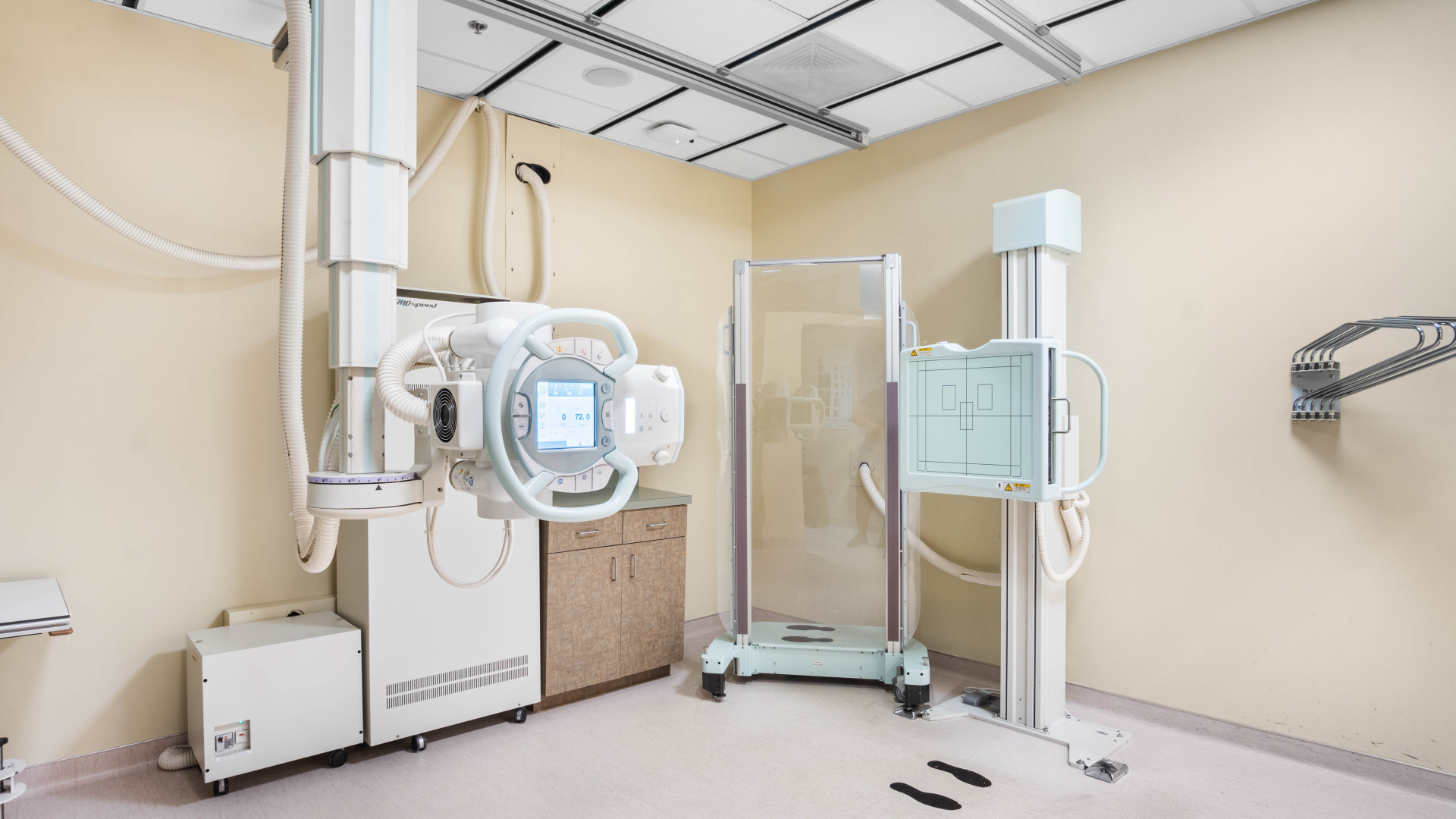
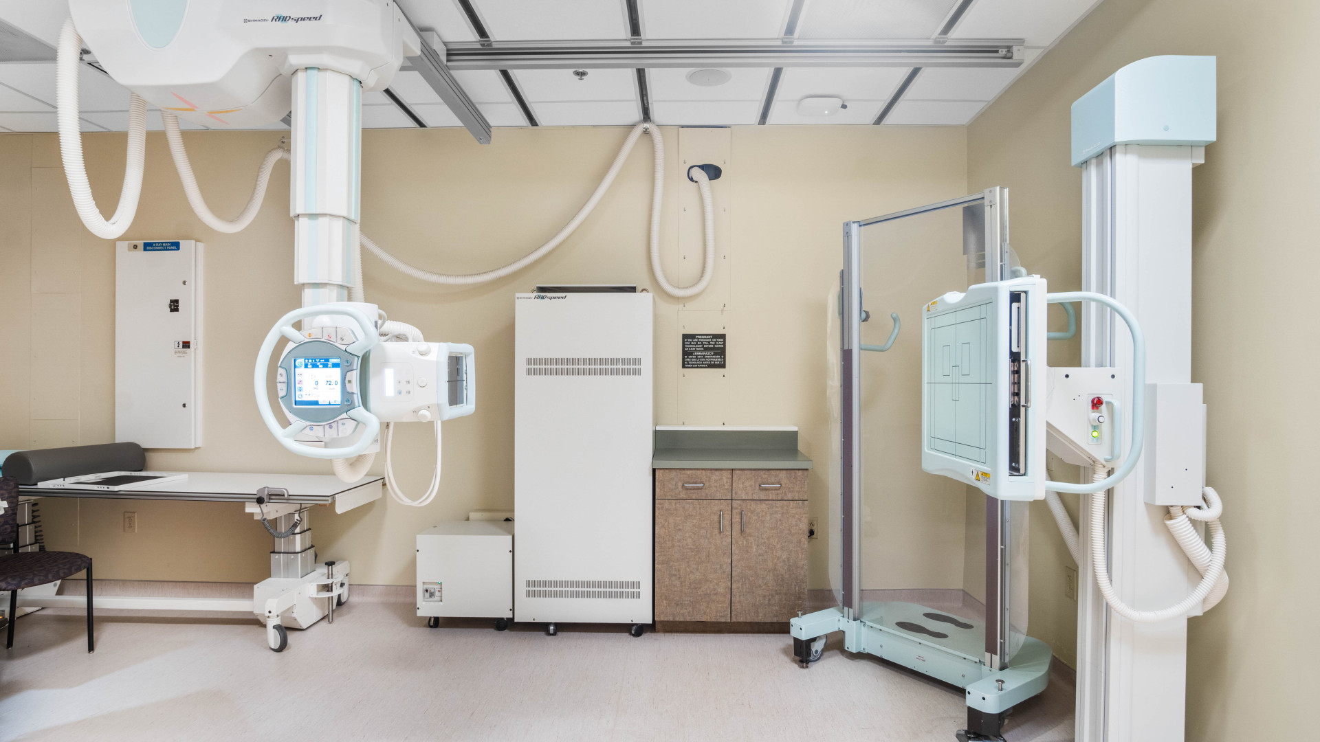
X-Ray
Imaging Services at Carolina Neurosurgery & Spine Associates
-
X-rays are a common and essential imaging technique widely used in
medical diagnostics. They are instrumental in:
- Detecting Fractures: X-rays can quickly and accurately reveal broken bones and fractures, making them indispensable in emergency and orthopedic medicine.
- Assessing Internal Organs: Beyond bones, X-rays can help visualize internal organs, such as the lungs and heart, to diagnose conditions like pneumonia, heart failure, and other chest conditions.
- Diagnosing Various Conditions: X-rays are used to identify a range of health issues, including infections, arthritis, bone tumors, and dental problems.
- Checking Surgical Hardware: Post-surgical X-rays are used to verify the correct placement and alignment of surgical hardware such as pins, plates, and screws.
- Spinal Alignment: X-rays are crucial for assessing the alignment and curvature of the spine, aiding in the diagnosis and treatment of conditions such as scoliosis, kyphosis, and other spinal disorders.
Injections with Guided Fluoroscopy
-
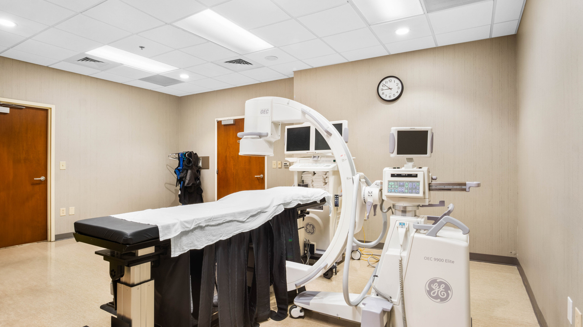
This advanced technique uses real-time X-ray imaging (fluoroscopy)
to precisely guide the placement of needles or catheters for various
medical procedures. It offers significant benefits in:
- Spine Procedures: Fluoroscopy is invaluable in guiding injections for pain management, such as epidural steroid injections and facet joint injections, ensuring accurate delivery of medication to the target area.
- Joint Procedures: It helps in administering injections into joints (e.g., shoulders, hips, knees) for pain relief or diagnostic purposes.
- Blood Vessel Procedures: Fluoroscopy aids in the accurate placement of catheters in blood vessels for treatments or diagnostic angiography.
- Medication Administration: Ensures that medications are delivered precisely to the intended location, enhancing the effectiveness and safety of the treatment.
- Convenience Patients can have their X-rays and practice appointments on the same day at one location.
- Consistency Highly skilled technologists perform every X-ray, ensuring tests are always consistent.
- Quick and Efficient X-ray imaging is fast, making it an excellent option for diagnosing fractures, infections, and other conditions quickly.


WHAT IS AN X-RAY?
X-rays are a form of electromagnetic
radiation that can pass through the body to create images of the
inside of the body. X-ray imaging is a quick, painless process that
helps doctors diagnose and monitor various medical conditions by
capturing detailed images of bones and certain internal organs.
HOW DOES A PATIENT PREPARE FOR AN X-RAY?
In most cases,
no special preparation is needed for an X-ray. You may be asked to
remove jewelry, glasses, and any other metal objects that could
interfere with the imaging. It's also advisable to wear loose-fitting
clothing. Depending on the area being examined, you might be asked to
wear a hospital gown. Always inform your doctor or X-ray technologist
if you are pregnant or suspect you might be.
HOW IS AN X-RAY PERFORMED?
You will be positioned by a
technologist in such a way that the part of the body being examined
is between the X-ray machine and the X-ray film or detector. You may
be asked to hold your breath for a few seconds to avoid movement that
could blur the image. The technologist will operate the X-ray machine
from behind a protective barrier or from an adjacent room to minimize
their exposure to radiation.
HOW LONG DOES THE TEST TAKE?
The actual X-ray exposure
typically takes less than a second, although the entire process,
including preparation and positioning, may take up to 15 minutes. If
multiple X-rays are needed, the process may take longer.
OTHER ITEMS TO CONSIDER
Notify your physician and the
technologist if you are pregnant, as X-ray radiation can be harmful
to an unborn baby. Inform them if you have any metal implants, as
these can interfere with the imaging process.


